Dermoid cysts teratomas - These complex ovarian cysts or mass are formed from the eggs produced and are often filled with pieces of bone teeth hair and cartilage. Hey all Ive been told I have a large solid mass covering my left ovary 81 x 60 x 76mm following an urgent ultrasound.
 A Left Ovarian Mass During Operation The Surface Is Smooth And The Download Scientific Diagram
Thats why its important to have regular pelvic exams.

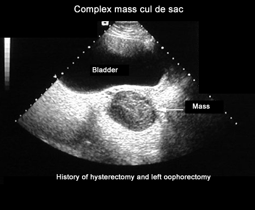
Mass on left ovary. Subacute hemorrhagic ovarian cyst simulating a solid ovarian mass. Options for treatment can range from surgery to birth control pills from chemotherapy to narcotic pain killers. The majority of ovarian cysts in pre-menopausal women will be normal cysts related to development of the egg in the ovary and ovulation.
An adnexal mass is a growth that occurs in or near the uterus ovaries fallopian tubes and the connecting tissues. The notes say with suspicious features of malignancy so clearly Im thinking the worst as it doesnt sound promising. Its rare but ovarian cysts.
One of the causes of pain in the left ovary is the pelvic inflammatory disease PID. The vast majority of ovarian neoplasms in girls and young women are not cancerous. Ovaries can become enlarged masses or tumors due to cysts ovarian and hemorrhagic ovarian cysts masses endometriomas or neoplasms growths.
Benign ovarian masses include functional cysts and tumors. This infection transfers during sexual intercourse such as chlamydia or gonorrhea. Ovarian tumors can be categorized as epithelial germ cell sex cordstromal or metastatic.
When they did the ultrasound they did not seem to know what it was ie. An ovarian cyst is a common condition which is typically not cancerous. If a cystic mass is cancerous your doctor will likely refer you to a gynecologic cancer specialist.
The ovaries are female reproductive organs that produce female hormones including estrogen. Tumors which occur in many areas of the body are abnormal growths that dont have any purpose. A mass was discovered on my left ovary.
They are usually benign and may grow to six inches in diameter. There are three types of complex ovarian cysts or mass and these are. CA125 slightly raised at 43.
Cystic ovarian masses that develop after menopause might be cancerous malignant. Your doctor is also likely to recommend surgery when an ovarian cyst develops after menopause. An ovarian mass refers to any lump cyst or tumor in the ovaries.
Endometriomas form when cells from your uterine lining grow outside of your uterus and in or on your ovaries. Fibroid or some unknown mass. Our Approach to Ovarian Masses.
To clear up one of your fears anyway it is very common for physicians to be perplexed by a pelvic mass in the region of the ovaries called an adnexal mass if you hear that term. A luteal cystic neoplasm or cyst of the yellow body of the left ovary is formed from the yellow body in the cortical layer of the ovary. You might need to have your uterus ovaries and fallopian tubes removed total hysterectomy and possibly chemotherapy or radiation.
Patients can be seen by Texas Childrens experts in Pediatric and Adolescent Gynecology. Infrequent complications associated with ovarian cysts include. An ovarian mass refers to an enlargement of one of the ovaries.
Epithelial tumors are the most common histopathologic type of malignant ovarian tumor 85 of cases Fig 1 5 6. The treatment for an ovarian mass will depend on the cause. An ovarian cyst is a fluid-filled sac in or on the ovary.
Cystadenomas contain ovarian tissue with fluid or mucus. In addition it can be a combination of a solid tumor and cyst. A tumor can be benign or cancerous malignant but ovarian tumors are typically benign.
Treating Ovarian Masses and Tumors. Cysts that enlarge can cause the ovary to move increasing the chance of painful twisting of your ovary ovarian torsion. Ovarian masses are growths on or in the ovaries the small reproductive organs located on each side of a girls uterus that store and release eggs and produces female hormones.
Subtypes of epithelial tumors include serous mucinous endometrioid clear cell and Brenner tumors. These cysts will almost always go away over time. Treatment varies depending on the patients reproductive status.
The corpus luteum is endocrine cells left over from a bursting follicle producing progesterone and dying off as the new ovulation approaches. Dual screen image without and with color Doppler interrogation shows a heterogeneous predominantly isoechoic solid-appearing mass between arrows within the left ovary of a 41-year-old woman who presented with pelvic pain. Causes Risk Factors.
Theyre usually benign but are sometimes cancerous. At the same time that doctors may not have any idea if an adnexal mass in your pelvis could be cancer the chance of being wrongacting before getting a better ideais equally concerning. A tumor is a solid mass unlike a fluid-filled ovarian cyst.
Men struggling with digestive neurological skeletal inflammatory prostate health issues complain of it. A healthy ovary and one with cysts. Typically an ovarian mass can present as a solid tumor or fluid-filled cyst.
An ovarian tumor is a slow-growing abnormal mass of tissue on or in a womans ovary. Blood work was done and I have an appointment on Monday. Some cysts and masses in or on the ovaries are benign while others can be cancerous and life-threatening.
Have been out on the 2ww for a gynae appt. Some of them are filled.
A cyst may become large enough to obscure the ovary from which it is arising. Radiology report The Society of Radiologists in Ultrasound made in 2019 the following recommendations regarding reporting of simple adnexal cysts of suspected ovarian origin based on size and menopausal status 2.
Ovarian cysts are.
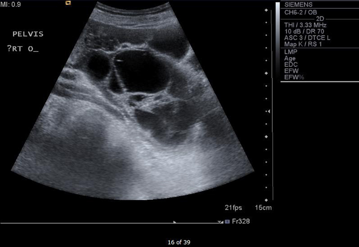
Large ovary on ultrasound. This means that the ovaries are small and not likely to be cancerous. If your ultrasound technician informs you that she cant see or find one of your ovaries do NOT panic or worry. The right ovary in this middle aged female patient see ultrasound images above shows a large primarily cystic mass with multiple septae.
Ovulation happens when these cysts are around 2 to 3 cm in size. These slow-growing tumors contain elements from multiple germ cell layers and are best assessed with ultrasound. Ovary ultrasound education showing how to scanning protocol normal anatomy anatomic variants follicles and graafian follicle corpus luteum.
An ultrasound uses sound waves to create images of the. Vaginal ultrasound can help to show whether any cysts on your ovaries contain cancer or not. Most of the time an enlarged ovary is a symptom of ovarian cysts polycystic ovary syndrome or ovarian cancer.
If cysts are large enough to cause symptoms like pain and bloating. Ovulation is a completely normal and healthy body function. Ovarian torsion happens when the ovary and part of the fallopian tube get twisted.
Your doctor can do an ultrasound or other imaging scans to see if your tumor has receded. In size and part of it shows coarse particulate matter producing fine echoes within the fluid. Sometimes in women who are past their menopause the ovaries do not show up on an ultrasound.
A wandlike device transducer sends and receives high-frequency sound waves ultrasound to create an image of your uterus and ovaries on a video screen. The cystic neoplasmmeasures 14 x 7 cms. ObjectiveTo evaluate calcifications5mminlengthin ovaries that are otherwise normal on ultrasound and to determine whether such large ovarian calcifications are an indicator of ovarian neoplasm.
However some may reach sizes of 8 to. Another method to locate the ovary is to trace along the external iliac vessels in a transverse section of the pelvis. Large ovarian calcifications seem to be encountered less frequently on sonography than are the more common 13mm echogenic foci 1 - 3 that have been the subject of most prior reports.
When the ovary releases the egg ovulation occurs. Ovulation can cause a change or increase in vaginal discharge and slight cramping. Ovarian dermoid cyst and mature cystic ovarian teratoma are terms often used interchangeably to refer to the most common ovarian neoplasm.
Calcifications can occur occasionally in various ovarian neoplasms. Doctors usually check the shape and size of the ovaries during a womans yearly pelvic examination. The complex solid-cystic lesions in addition to being bilateral are suspicious for a cystic ovarian neoplasm and warrant further evaluation.
The ultrasound technician told me that she was unable to see my left ovary. This usually happens around day 14 of a 28-day cycle. If a cyst has any solid areas it is more likely to be cancer.
Ovarian cysts are sacs filled with fluid that can form inside or outside the ovaries. The ovaries can usually be located on ultrasound by angulating the probe from a TS view of the uterus along the hypoechoic utero-ovarian ligament from one cornu to the lateral pelvic wall. The ultrasound technician gave me a very normal reason why she could not find my left ovary.
These appearances are typical of mucinous cystadenoma of the right ovary. On ultrasound both ovaries are markedly enlarged and contain cystic components with intracystic solid components arrows. Otherwise polycystic ovaries can be considered a normal variant.
Echogenic foci 5mm and larger some of which may demonstrate posterior acoustic shadowing seen in otherwise normal ovaries have been attributed to corpus albicans. Most functional cysts are 2 to 5 centimeters cm about 34 of an inch to 2 inches in size. Polycystic ovaries PCO or polycystic ovarian morphology is an imaging descriptor of a particular type of change in ovarian morphologyA proportion of women with polycystic ovaries will have the polycystic ovarian syndrome PCOS which in turn requires additional clinical as well as biochemical criteria.
Your doctor analyzes the image to confirm the presence of a cyst help identify its location and determine whether its solid filled with fluid or mixed. If she says its hidden do NOT worry. When someone has symptoms that could indicate enlarged ovaries or another ovarian condition a doctor is likely to recommend an ultrasound.
Larger echogenic foci in the ovary usually from isolated calcifications are also typically benign findings Fig.