Thyroid nodules can be detected in 4 to 8 of the adult population by palpation but in 40 to 50 of the population by ultrasound. Ultrasound uses soundwaves to create a picture of the structure of the thyroid gland and accurately identify and characterize nodules within the thyroid.
 Ultrasonographic Features For Differentiating Follicular Thyroid Carcinoma And Follicular Adenoma Sciencedirect
Imaging techniques particularly high resolution sonography of the neck have been shown to.

Malignant thyroid nodule ultrasound. Malignancy prevalence in nodules with a highly suspicious ultrasound category based on ATA Criteria was 685 significantly higher than indeterminate nodules with low-risk ultrasound patterns per the ATA. 1 They are palpable in 47 of the population and have been detected using ultrasonography in up to 67 of adults. The ACR-TIRAD uses ultrasound data and a point system based on nodule composition echogenicity nodule fluid content shape nodule margins and echogenic foci particulates within the nodule of differing echogenicity 6-8.
Since thyroid nodules are frequently detected by cervical ultrasound examinations distinguishing between a benign and a malignant nodule is a relevant clinical challenge. Clinical Oncology Feb 21 2017. Thyroid nodules are commonly detected on physical examination and even more commonly identified as incidental findings on computed tomography CT magnetic resonance imaging MRI radionuclide studies and ultrasound examinations of the neck done for other purposes than evaluating the thyroid gland Fig.
Similar results were found when researchers stratified for papillary thyroid carcinoma and its variants. Ultrasound is the first-line imaging modality for assessment of thyroid nodules found on clinical examination or incidentally on another imaging modality. Suspiciously malignant findings on ultrasound after fine needle aspiration biopsy in a thyroid nodule with initially benign ultrasound and cytologic result.
The overwhelming majority of these represent benign hyperplastic nodules or adenomas. 2 While the majority of nodules are benign the risk of malignancy reaches approximately 715. Approximately 5 of nodules are malignant with papillary carcinoma representing approximately 75.
Ultrasound of Thyroid Nodules. Thyroid nodules are discrete lesions present within the thyroid gland that are radiologically distinct from the adjacent parenchyma Table 1. Zhang eet al Int J.
A few Radiomics studies have been conducted on ultrasound images for classifying benign and malignant thyroid nodules 91821. To repeat or to follow-up Clinical Imaging Vol. A simple procedure that is done in the doctors office to determine if a thyroid nodule is benign.
Partially cystic thyroid nodules on ultrasound. The total point value is divided into five levels with TR1 and TR2 being the lowest. Ultrasound USG can be a good screening tool to identify high-risk nodule requiring fine-needle aspiration cytology FNAC.
A cross-sectional study was performed from August 2011 to July 2012 at Tribhuvan University Teaching Hospital. 1 Given the prevalence of thyroid nodules and their underlying risk it is imperative that general practitioners GPs are able to. Thyroid malignancy is relatively rare and is diagnosed in approximately 25000 patients per year in the United States 1.
Thyroid fine needle aspiration biopsy FNAB. Height of the nodule nodular calcifications ill defined nodule borders irregular shape were associated with a higher score for malignancy. Thyroid nodules are commonly noted in adults.
This article is an overview of ultrasonographic features of thyroid nodules which are used to determine the need for biopsy with fine needle aspiration. The prevalence of palpable thyroid nodules is estimated to be 64 in women and 15 in men between 30 to 60 years of age living in iodine-sufficient regions. The most common cause of benign thyroid nodules is nodular hyperplasia 2.
With respect to current guidelines it is the sonographic pattern rather than the growth of a nodule that raises suspicions of malignancy 15. Probability of malignancy and sonographic differentiation About 5 of partially cystic nodules in our series were malignant. A common imaging test used to evaluate the structure of the thyroid gland.
The study aimed to assess the association of USG characteristic of thyroid nodule with malignancy. When more than 50 of the nodule is solid and the solid portion of the nodule is eccentric the risk of malignancy is greater. Thyroid nodules are a common clinical finding and are increasingly detected in the general population due to the widespread use of thyroid ultrasound US examination 1 2Fine-needle aspiration FNA biopsy is currently the main diagnostic tool for the detection of the minority of thyroid lesions that result to be malignant 3 4Its use should be restricted to thyroid nodules associated.
Ultrasound can detect the presence site size and number of thyroid nodules and there have been reports of US characteristics of malignancy such as ill-defined margin irregular shape hypoechogenicity heterogeneity absence of cystic lesion andor the halo sign the presence of calcification and invasion to adjacent organs. Ultrasound is also frequently used to guide the needle into a nodule during a thyroid nodule biopsy. Also the study suggested a less than 2 cm under ultrasonography as the cutoff for malignancy.
Ultrasound can help evaluate a thyroid nodule and determine the need for biopsy. A thyroid fine needle aspiration biopsy can collect samples of cells from the nodule which under a microscope can provide your doctor with more information about the behavior of the nodule. What does a Malignant Thyroid Nodule Look Like on Ultrasound Imaging.
Therefore it is worthy of investigating whether a Radiomics approach can make better use of thyroid ultrasound images and achieve more accurate diagnosis of differentiating malignant from benign thyroid nodules. Ultrasound-guided fine-needle aspiration cytopathology can predict oversize of thyroid nodule than original but may predict the risk of malignancy of the thyroid nodule. Whats the treatment for a thyroid nodule.
Thyroid nodules are very common and may be observed at ultrasonography US in 50 of the adult population.
Gain Focus Harmonic Denoise. Convex 100mm280mm Linear 20mm55mm.
 Wireless Ios Fetal Doppler Convex Probe Veterinary Ultrasound Keebovet Veterinary Ultrasound Equipment
32 out of 5 stars 6.

Ultrasound doppler for iphone. IOS and Android compatibleAffordable Price For clinical home usePaypal accept. IPhone Apps as Doppler. Mobisante an mHealth company based in Redmond WA has recently been showing a new smartphone peripheral at conferences across the country an ultrasound probe.
Premium image quality from a portable handheld ultrasound scanner you can carry anywhere Class-Leading Image Quality Wireless App Based Ultrasound for iPhone Android. Discussion in Pregnancy - Second Trimester started by KatieKoopa May 7 2011. So I suspect its a load of utter shit.
A wide variety of iphone ultrasound scanner options are available to you such as ce ccc. One such innovation is called the Butterfly iQ a small ultrasound device that can display clear black-and-white pictures on an iPhone. Handheld ultrasound u-lite an advanced diagnostic tool U-Lite is always ready to go further than a simple palpation.
Ultrasound Portable Iphone Manufacturers Factory Suppliers From China Welcome to go to us anytime for company partnership proven. Download Butterfly iQ Ultrasound and enjoy it on your iPhone iPad and iPod touch. Online Store for Portable handheld wireless wifi ultrasound b probe scannerdisplay on iphoneipadTabletsSmartphoneslaptop.
Connect to PC Tablets Smartphones. Wonderful clarity of images in a very intuitive interface. SonoQue is a portable doppler scanner.
370 Goddard Irvine CA 92618. For love image the world. However it is useful for quick ultrasound diagnosis.
A big boon for the bedside ultrasound community. We tried a iphone doppler thing to check the babys heart beat jusy for fun but there was nothing everything was fine last week at the midwives appointment but should i be worried. Connect the Lumify transducer Our innovation in portable ultrasound comes to your compatible device through a simple USB or Lightning connection.
IOS and Android compatible. Easy to use this handheld ultrasound scanner is entirely dedicated to help health professionals make high-quality care and diagnostic. It just uses the iphones mic to try and pick the hb up and amplify it.
Do they actually work. You can also choose from plastic metal and steel iphone ultrasound scanner as well as from 2 years 1 year and lifetime iphone ultrasound scanner and whether iphone ultrasound scanner is online technical support onsite training or none. These include the incident frequency of the ultrasound beam used speed of.
Designed as a Point-and-Shoot Ultrasound system it uses advanced AI technologies to instantly adjust settings for a great image you can trust. SFMT Ultrasound Wireless Doppler is complete and accurate like a traditional Doppler but with the advantage of being portable. However several factors influence the resultant frequency shift and hence the measured velocity.
The price is very inexpensive and you can use ultrasound easily but the resolution is downside compare to the advanced doppler. The robust Lumify handheld ultrasound app is available on compatible Android and Apple mobile devices via the Google Play Store and the Apple App store. DW-P5 3d 4d color doppler ultrasound machine.
Good for home clinics vet. It works with every iOS Android and Windows device. Connect your Clarius HD ultrasound scanner to your phone or tablet with this easy-to-use Clarius App.
According to the MIT Technology Review the current prototype connects to a Toshiba TG01 smartphone was originally developed as a laptop peripheral by David Zar a computer. This concept of a Doppler shift is used to measure blood flow velocity whereby the shift in sound-wave frequency is detected by the ultrasound transducer after encountering active blood flow. Widely clinical applications meets daily diagnosis.
Is anyone using any apps as a doppler. Mar 5 2011 Messages. Clarius HD delivers the highest definition ultrasound image in a wireless handheld scanner.
Wireless Wifi Convex Array Probe Type Ultrasound Scanner 35Mhz128E. Students love it residents are begging for it and faculty are demanding more access to this technology. Iphone us venous color doppler scan MSLPU55 Specification.
B BM and Color PW PDI. More DW-F5 economical type 3d4d doppler ultrasound b. Ultrasound technology has evolved rapidly over recent decades.
Its not an ultrasound Doppler like a handheld one. Butterfly iQ provides whole body ultrasound imaging with just one probe. 19 clinical presets and familiar touch interactions for unprecedented ease of use.
How to use is very simple and easy. 75 MHz100MHz and 10MHz14MHz -Depth. How far along are you.
This will change the game of mobile ultrasound market in terms of quality portability connectivity interface and cost. The L5C wireless portable color doppler linear transducer probe machine 75 - 10 MHz brings diagnostics to your iPhone iPad. Page 1 of 3 1 2 3 Next KatieKoopa Mama of 1 Pregs with 2.
A cyst may become large enough to obscure the ovary from which it is arising. Radiology report The Society of Radiologists in Ultrasound made in 2019 the following recommendations regarding reporting of simple adnexal cysts of suspected ovarian origin based on size and menopausal status 2.
Ovarian cysts are.
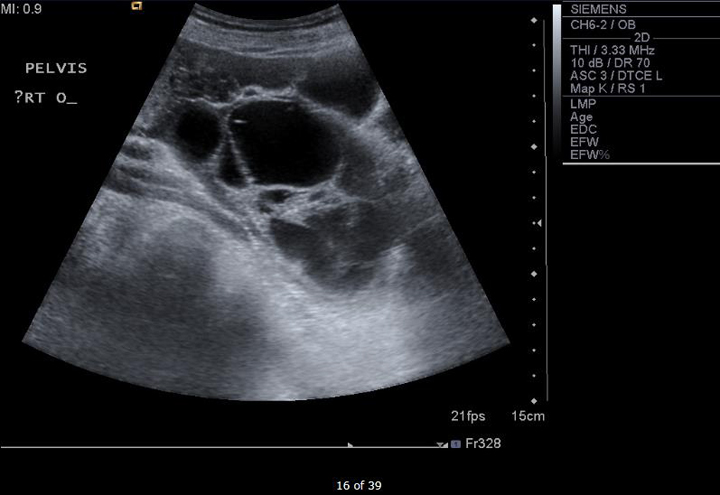
Large ovary on ultrasound. This means that the ovaries are small and not likely to be cancerous. If your ultrasound technician informs you that she cant see or find one of your ovaries do NOT panic or worry. The right ovary in this middle aged female patient see ultrasound images above shows a large primarily cystic mass with multiple septae.
Ovulation happens when these cysts are around 2 to 3 cm in size. These slow-growing tumors contain elements from multiple germ cell layers and are best assessed with ultrasound. Ovary ultrasound education showing how to scanning protocol normal anatomy anatomic variants follicles and graafian follicle corpus luteum.
An ultrasound uses sound waves to create images of the. Vaginal ultrasound can help to show whether any cysts on your ovaries contain cancer or not. Most of the time an enlarged ovary is a symptom of ovarian cysts polycystic ovary syndrome or ovarian cancer.
If cysts are large enough to cause symptoms like pain and bloating. Ovulation is a completely normal and healthy body function. Ovarian torsion happens when the ovary and part of the fallopian tube get twisted.
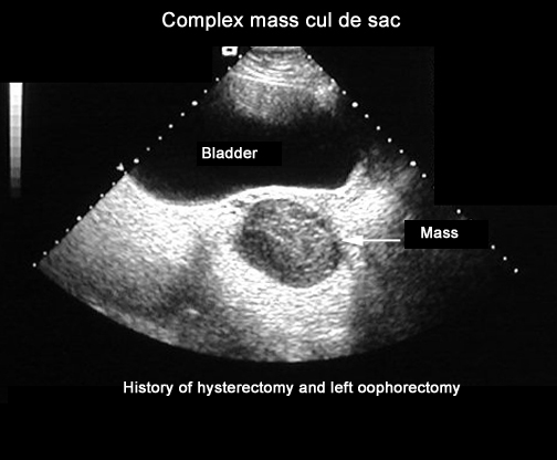
Your doctor can do an ultrasound or other imaging scans to see if your tumor has receded. In size and part of it shows coarse particulate matter producing fine echoes within the fluid. Sometimes in women who are past their menopause the ovaries do not show up on an ultrasound.
A wandlike device transducer sends and receives high-frequency sound waves ultrasound to create an image of your uterus and ovaries on a video screen. The cystic neoplasmmeasures 14 x 7 cms. ObjectiveTo evaluate calcifications5mminlengthin ovaries that are otherwise normal on ultrasound and to determine whether such large ovarian calcifications are an indicator of ovarian neoplasm.
However some may reach sizes of 8 to. Another method to locate the ovary is to trace along the external iliac vessels in a transverse section of the pelvis. Large ovarian calcifications seem to be encountered less frequently on sonography than are the more common 13mm echogenic foci 1 - 3 that have been the subject of most prior reports.
When the ovary releases the egg ovulation occurs. Ovulation can cause a change or increase in vaginal discharge and slight cramping. Ovarian dermoid cyst and mature cystic ovarian teratoma are terms often used interchangeably to refer to the most common ovarian neoplasm.
Calcifications can occur occasionally in various ovarian neoplasms. Doctors usually check the shape and size of the ovaries during a womans yearly pelvic examination. The complex solid-cystic lesions in addition to being bilateral are suspicious for a cystic ovarian neoplasm and warrant further evaluation.
The ultrasound technician told me that she was unable to see my left ovary. This usually happens around day 14 of a 28-day cycle. If a cyst has any solid areas it is more likely to be cancer.
Ovarian cysts are sacs filled with fluid that can form inside or outside the ovaries. The ovaries can usually be located on ultrasound by angulating the probe from a TS view of the uterus along the hypoechoic utero-ovarian ligament from one cornu to the lateral pelvic wall. The ultrasound technician gave me a very normal reason why she could not find my left ovary.
These appearances are typical of mucinous cystadenoma of the right ovary. On ultrasound both ovaries are markedly enlarged and contain cystic components with intracystic solid components arrows. Otherwise polycystic ovaries can be considered a normal variant.
Echogenic foci 5mm and larger some of which may demonstrate posterior acoustic shadowing seen in otherwise normal ovaries have been attributed to corpus albicans. Most functional cysts are 2 to 5 centimeters cm about 34 of an inch to 2 inches in size. Polycystic ovaries PCO or polycystic ovarian morphology is an imaging descriptor of a particular type of change in ovarian morphologyA proportion of women with polycystic ovaries will have the polycystic ovarian syndrome PCOS which in turn requires additional clinical as well as biochemical criteria.
Your doctor analyzes the image to confirm the presence of a cyst help identify its location and determine whether its solid filled with fluid or mixed. If she says its hidden do NOT worry. When someone has symptoms that could indicate enlarged ovaries or another ovarian condition a doctor is likely to recommend an ultrasound.
Larger echogenic foci in the ovary usually from isolated calcifications are also typically benign findings Fig.